We moved to the farm in November 2008. As city kids we were absolutely thrilled to have some land for gardening and even a little livestock... Naturally, we went nuts! I had a huge garden the following year, and this place is blessed with loads of prolific if elderly fruit trees, so I canned and canned and canned. The year after, 2010, I attempted very little in terms of gardening because we just had too much going on.
But the one thing we've stuck to all along even when I couldn't manage any gardening is... chickens!
We got our first chicks in early spring 2009. Plodding steadily on, we tried many things, all aiming at a simple goal of a sustainable supply of chicken meat and eggs. A laying flock is easy, but meat is a little more challenging as I don't want to buy chicks from hatcheries or feed stores. On the other hand growing my own grains is far too ambitious a project for me. So in my ideal world, we will be breeding all our own chicks and we will be able to sell enough eggs (and maybe chickens) to pay for the cost of feeding the flocks. These are pastured flocks, by the way, that are doing quite a bit of foraging too.
At last we're getting close! I have a freezer full of chicken and we haven't bought eggs in at least a year.
I have a laying flock of black Australorps.
I think they're beautiful as well as great dual-purpose birds... strong layers and great meat in a reasonable time. I've worked out a deal with a farming friend who started her own hatchery last year but has no Australorp stock. So now I collect hatching eggs now and then, she hatches them along with all her others, and then we split the 'Lorp chicks 50/50. So of the chicks I get, when they grow up, the girls join the laying flock and the boys go into the freezer. Another farming friend nearby comes here with a plucker on the back of her truck and does all the butchering.
Of course most people raising meat birds go with cornish cross, which mature much faster so are cheaper to raise, but those things are the opposite of sustainable. They can't reproduce naturally, and they can't forage. The poor things are engineered to be little eating machines that will drop dead of heart attacks if you don't butcher them on time. Not for me. On the other hand I can't afford to be raising gourmet birds. Hence the sustainability goal where cash from selling eggs offsets extra feed costs.
Meantime, my daughter Chaidie is doing a great job taking care of her own laying flock, which is a miscellany of colored egg layers (Americaunas), some "leftover" barred rocks from a batch of birds we raised a year or so ago, some decorative birds (white and golden Polish that we raised; the sole Houdan survivor after a bobcat started visiting my sister's free-range buffet; and a beautiful English game bantam rooster I accidentally acquired by foolishly checking the "Please send me a free exotic chick" box while ordering a batch of birds online from a hatchery once), plus various foster chickens who were driven out of their homes by a neighboring city's prejudice against backyard poultry.
When we give kitchen scraps to the birds, the barn cats sometimes get jealous...
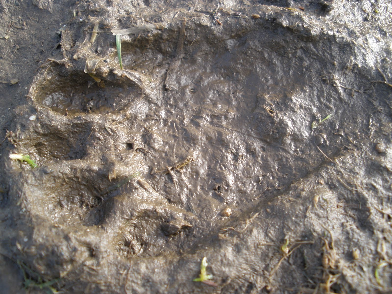
This fall/winter have brought a lot of hyperactive wildlife to our neighborhood. Here's a track in our driveway:
Thursday, January 27, 2011
Abstract: So when you walk out into the bracing cold...
...Your eyes get drier but they still water. Another of the perennial counterintuitive parts of what is more appropriately, though not quite so handily, called dysfunctional tear syndrome. Hand me my Wileys, will you?
Ocular surface wetness is regulated by TRPM8-dependent cold thermoreceptors of the cornea.
Nat Med. 2010 Dec;16(12):1396-9. Epub 2010 Nov 14.
Parra A, Madrid R, Echevarria D, del Olmo S, Morenilla-Palao C, Acosta MC, Gallar J, Dhaka A, Viana F, Belmonte C.
Instituto de Neurociencias de Alicante, Universidad Miguel Hernández-Consejo Superior de Investigaciones Científicas, San Juan de Alicante, Spain.
Ocular surface wetness is regulated by TRPM8-dependent cold thermoreceptors of the cornea.
Basal tearing is crucial to maintaining ocular surface wetness. Corneal cold thermoreceptors sense small oscillations in ambient temperature and change their discharge accordingly. Deletion of the cold-transducing ion channel Transient receptor potential cation channel subfamily M member 8 (TRPM8) in mice abrogates cold responsiveness and reduces basal tearing without affecting nociceptor-mediated irritative tearing. Warming of the cornea in humans also decreases tearing rate. These findings indicate that TRPM8-dependent impulse activity in corneal cold receptors contributes to regulating basal tear flow.
Nat Med. 2010 Dec;16(12):1396-9. Epub 2010 Nov 14.
Parra A, Madrid R, Echevarria D, del Olmo S, Morenilla-Palao C, Acosta MC, Gallar J, Dhaka A, Viana F, Belmonte C.
Instituto de Neurociencias de Alicante, Universidad Miguel Hernández-Consejo Superior de Investigaciones Científicas, San Juan de Alicante, Spain.
Wednesday, January 26, 2011
Abstract: Why your eyes don't sparkle the way they used to
Of all the things that bother me about dry eye, I have to say this is pretty low on my list...
The Sparkle of the Eye: The Impact of Ocular Surface Wetness on Corneal Light Reflection.
Am J Ophthalmol. 2011 Jan 20. [Epub ahead of print]
Goto E, Dogru M, Sato EA, Matsumoto Y, Takano Y, Tsubota K.
Department of Ophthalmology, School of Dental Medicine, Tsurumi University, Yokohama, Japan; Department of Ophthalmology, School of Medicine, Keio University, Tokyo, Japan.
The Sparkle of the Eye: The Impact of Ocular Surface Wetness on Corneal Light Reflection.
PURPOSE: To measure the sparkle of the human eye evaluated by the intensity of corneal light reflection in normal subjects and dry eye patients to investigate whether ocular surface wetness has an impact on the sparkle of the eye.
DESIGN: Prospective case-control study.
METHODS: We examined a consecutive series of eight dry eye patients with Sjögren syndrome (SS, 15 eyes), as well as eight normal subjects (16 eyes). The specular corneal surface light reflection was quantitatively measured with an ophthalmic slit-lamp microscope and image capturing system under fixed conditions of light source, incident angle, and detector sensitivity. The intensity of images from subjects' corneal light reflection was quantified with image analysis software along with the measurement of grade of self-reported brilliancy of the eye, corneal fluorescein staining score, tear film break-up time, and Schirmer test value. The intensity of corneal light reflection was also compared before and after dry eye treatment.
RESULTS: The mean intensity of corneal light reflection was significantly lower in dry eye patients (125.0 ± 40.1) than normal subjects (167.6 ± 36.6, P = .004). Grade of self-reported brilliancy of the eye, corneal fluorescein staining scores, tear film break-up time, and Schirmer test values showed good correlation to the intensity of corneal light reflection. After punctal plug treatment, the intensity of corneal light reflection significantly increased from 125.0 ± 40.1 to 167.2 ± 45.0 (P < .0001).
CONCLUSIONS: The intensity of corneal light reflection representing the sparkle of the eye was significantly more intense in normal subjects compared to dry eye patients, and was increased after punctal plug treatment. The intensity of corneal light reflection appeared to correlate well with tear film stability, volume, and ocular surface desiccation. We showed that tears contributed not only to ocular surface wetness but also to the extent of the light reflection from the eye.
Am J Ophthalmol. 2011 Jan 20. [Epub ahead of print]
Goto E, Dogru M, Sato EA, Matsumoto Y, Takano Y, Tsubota K.
Department of Ophthalmology, School of Dental Medicine, Tsurumi University, Yokohama, Japan; Department of Ophthalmology, School of Medicine, Keio University, Tokyo, Japan.
Abstract: Relationship between clinical signs and dry eye symptoms
Talk about a charged subject. This sounded quite interesting though and the results are actually modest enough to be believable (as opposed to looky, my new tool correlates just great with symptoms!)
The relationship between clinical signs and dry eye symptoms.
Eye (Lond). 2011 Jan 21. [Epub ahead of print]
Pult H, Purslow C, Murphy PJ.
1] Optometry and Vision Research, Weinheim, Germany [2] School of Optometry and Vision Sciences, Contact Lens and Anterior Eye Research (CLAER) Unit, Cardiff University, Wales, UK.
The relationship between clinical signs and dry eye symptoms.
Purpose
To evaluate (i) the relationship between traditional and new clinical tests (lid-wiper epitheliopathy (LWE), lid-parallel conjunctival folds (LIPCOF)) and dry eye symptoms in non-contact lens wearers, and (ii) that a combination of these tests can improve predictive ability for the development of dry eye symptoms.
Methods
Tear meniscus height (TMH), non-invasive break-up time (NIBUT), ocular hyperaemia, LIPCOF, phenol red thread test (PRTT), corneal and conjunctival staining, and LWE grades were observed in a cohort of 47 healthy, non-lens wearers (male=17, female=30, median age=35 years, range=19-70). Symptoms were assessed using the Ocular Surface Disease Index (OSDI).
Results
LWE was significantly correlated to both temporal and nasal LIPCOF (0.537 < r < 0.607, P < 0.05). LIPCOF and LWE were significantly correlated to NIBUT and PRTT (r>-0.248, P<0.001). Significant correlations were found between NIBUT and TMH (r=0.461, P=0.001) and PRTT (r=0.640, P<0.001). OSDI scores were significant correlated to NIBUT, TMH, PRTT, LIPCOF, and LWE (r>∣0.31∣; P<0.05). Significant discriminators of OSDI+/- were NIBUT (area under the receiver operative characteristic curve (AUC)=0.895), TMH (0.715), PRTT (0.781), LIPCOF (temporal/nasal/Sum 0.748/0.828/0.816), and LWE (0.749). Best predictive ability was achieved by combining NIBUT with nasal LIPCOF (AUC=0.944).
Conclusions
The individual tests NIBUT, TMH, PRTT, LIPCOF, and LWE were significantly, but moderately, related to OSDI scores. The strongest relationship appeared by combining NIBUT with nasal LIPCOF.
Eye (Lond). 2011 Jan 21. [Epub ahead of print]
Pult H, Purslow C, Murphy PJ.
1] Optometry and Vision Research, Weinheim, Germany [2] School of Optometry and Vision Sciences, Contact Lens and Anterior Eye Research (CLAER) Unit, Cardiff University, Wales, UK.
Abstract: Phospholipid transfer protein
This was pretty interesting. But the main reason I'm posting it is in case anyone in the region (Finland & thereabouts) who's struggling to find a dry eye literature doctor might be able to scavenge useful names from the author list.
Interaction of phospholipid transfer protein with human tear fluid mucins.
Setälä NL, Holopainen JM, Metso J, Yohannes G, Hiidenhovi J, Andersson LC, Eriksson O, Robciuc A, Jauhiainen M.
J Lipid Res. 2010 Nov;51(11):3126-34. Epub 2010 Aug 19.
Department of Ophthalmology, University of Helsinki, Finland.
Interaction of phospholipid transfer protein with human tear fluid mucins.
In addition to circulation, where it transfers phospholipids between lipoprotein particles, phospholipid transfer protein (PLTP) was also identified as a component of normal tear fluid. The purpose of this study was to clarify the secretion route of tear fluid PLTP and elucidate possible interactions between PLTP and other tear fluid proteins. Human lacrimal gland samples were stained with monoclonal antibodies against PLTP. Heparin-Sepharose (H-S) affinity chromatography was used for specific PLTP binding, and coeluted proteins were identified with MALDI-TOF mass spectrometry or Western blot analysis. Immunoprecipitation assay and blotting with specific antibodies helped to identify and characterize PLTP-mucin interaction in tear fluid. Human tear fluid PLTP is secreted from the lacrimal gland. MALDI-TOF analysis of H-S fractions identified several candidate proteins, but protein-protein interaction assays revealed only ocular mucins as PLTP interaction partners. We suggest a dual role for PLTP in human tear fluid: (1) to scavenge lipophilic substances from ocular mucins and (2) to maintain the stability of the anterior tear lipid film. PLTP may also play a role in the development of ocular surface disease.
Setälä NL, Holopainen JM, Metso J, Yohannes G, Hiidenhovi J, Andersson LC, Eriksson O, Robciuc A, Jauhiainen M.
J Lipid Res. 2010 Nov;51(11):3126-34. Epub 2010 Aug 19.
Department of Ophthalmology, University of Helsinki, Finland.
Abstract: Phakic IOL & dry eye
This abstract covers exactly the kind of thing I have known must be going on but which I still dread seeing in print:
a) People getting phakic IOLs instead of LASIK/PRK because they have dry eye (and heaven forbid that someone should NOT be a candidate for some refractive surgery somewhere, especially in the military), PLUS
b) Nobody (apparently?) looking seriously at the ocular surface impact in the long run.
This latter point may sound like a stretch as regards the study whose abstract appears below. Honestly, I would be delighted to be contradicted with facts. If you can tell me that in this study, ocular surface assement methods which are now standard in OSD studies (OSDI, staining, TBUT and so on) were employed at 3 months postoperatively and NO PATIENTS had clinical signs or symptoms of dry eye at 3 months postop, I'll happily stand corrected. On the other hand if you tell me that "significant postoperative complications" is not intended to include dry eye - which is statistically the most frequently occurring complication of the more popular LASIK, and is disclosed as such in most informed consent forms - , I will be not the least bit surprised.
I hear from a ton of LASIK patients with dry eye because a ton of LASIK is done and a small percentage of the ones with dry eye end up on the internet. Very little phakic IOL is done (compared to LASIK) and I rarely hear from patients, but I have enough reports of dry eye after cataract surgery that I can't imagine it's not happening with elective lens implants.
Understand that I'm not bashing Phakic IOLs as such (I might but that's a different blog post). I just don't like any elective refractive surgery on already dry eyes, nor yet the tendency to assume it won't affect the ocular surface in healthy eyes.
Phakic Intraocular Lens Implantation in United States Military Warfighters: A Retrospective Analysis of Early Clinical Outcomes of the Visian ICL.
Darned tootin' further study is needed. But geez, IOLs have been around long enough. Why ain't we doing this yet? If it's been done where is it? I've had a MedLine feed on anything with dry eye in the abstract for years. [URL="http://www.journalofrefractivesurgery.com/"]JRS[/URL] scarcely ever even gives dry eye a cursory glance. Much to be expected, I guess.
J Refract Surg. 2011 Jan 17:1-9. doi: 10.3928/1081597X-20110106-03. [Epub ahead of print]
Parkhurst GD, Psolka M, Kezirian GM.
a) People getting phakic IOLs instead of LASIK/PRK because they have dry eye (and heaven forbid that someone should NOT be a candidate for some refractive surgery somewhere, especially in the military), PLUS
b) Nobody (apparently?) looking seriously at the ocular surface impact in the long run.
This latter point may sound like a stretch as regards the study whose abstract appears below. Honestly, I would be delighted to be contradicted with facts. If you can tell me that in this study, ocular surface assement methods which are now standard in OSD studies (OSDI, staining, TBUT and so on) were employed at 3 months postoperatively and NO PATIENTS had clinical signs or symptoms of dry eye at 3 months postop, I'll happily stand corrected. On the other hand if you tell me that "significant postoperative complications" is not intended to include dry eye - which is statistically the most frequently occurring complication of the more popular LASIK, and is disclosed as such in most informed consent forms - , I will be not the least bit surprised.
I hear from a ton of LASIK patients with dry eye because a ton of LASIK is done and a small percentage of the ones with dry eye end up on the internet. Very little phakic IOL is done (compared to LASIK) and I rarely hear from patients, but I have enough reports of dry eye after cataract surgery that I can't imagine it's not happening with elective lens implants.
Understand that I'm not bashing Phakic IOLs as such (I might but that's a different blog post). I just don't like any elective refractive surgery on already dry eyes, nor yet the tendency to assume it won't affect the ocular surface in healthy eyes.
Phakic Intraocular Lens Implantation in United States Military Warfighters: A Retrospective Analysis of Early Clinical Outcomes of the Visian ICL.
PURPOSE:
To assess short-term clinical outcomes after implantation of phakic intraocular lenses (Visian ICL, STAAR Surgical Co) in US military warfighters who are not good candidates for laser vision correction.
METHODS:
A retrospective interventional consecutive case series analysis of all eyes that underwent ICL surgery during a 14-month time period was performed. Main outcome measures included indications for surgery, efficacy, predictability, and early adverse events.
RESULTS:
Three-month postoperative visual data were available for 135 eyes of 69 patients who underwent ICL implantation during the study period. Indications included abnormal corneal topography (37%), thin predicted residual bed following LASIK (32%), history of dry eye (13%), thin corneal thickness (11%), or other (7%). Mean patient age was 30.9±6.6 years. Mean preoperative spherical equivalent refraction was -6.00±1.92 diopters (D) (range: -2.63 to -11.50 D). Three months postoperative, uncorrected distance visual acuity of 20/20 or better was found in 129/135 (96%) eyes and 91/135 (67%) were 20/15 or better. Manifest refraction and corrected distance visual acuity (CDVA) data were available for 128 eyes. Forty-two (33%) eyes had improvement of one or more lines of CDVA. One hundred fifteen eyes (90%) were within ±0.50 D of emmetropia, and predictability within ±0.75 D was found in 127/128 (99%) eyes. No significant intra- or postoperative complications were observed.
CONCLUSIONS:
This retrospective analysis of 3-month outcomes suggests that Visian ICL implantation in myopic warfighters provides excellent refractive and visual results. Further study is needed to evaluate long-term results.
Darned tootin' further study is needed. But geez, IOLs have been around long enough. Why ain't we doing this yet? If it's been done where is it? I've had a MedLine feed on anything with dry eye in the abstract for years. [URL="http://www.journalofrefractivesurgery.com/"]JRS[/URL] scarcely ever even gives dry eye a cursory glance. Much to be expected, I guess.
J Refract Surg. 2011 Jan 17:1-9. doi: 10.3928/1081597X-20110106-03. [Epub ahead of print]
Parkhurst GD, Psolka M, Kezirian GM.
Abstract: In vitro dry eye model
Very interesting, after all the mice and bunny models we usually seel.
Molecular mechanism of ocular surface damage: Application to an in vitro dry eye model on human corneal epithelium.
Mol Vis. 2011 Jan 12;17:113-26.
Meloni M, De Servi B, Marasco D, Del Prete S.
Molecular mechanism of ocular surface damage: Application to an in vitro dry eye model on human corneal epithelium.
PURPOSE:
The present study was concerned with the development of a new experimental model of dry eye using human reconstructed in vitro corneal epithelium (HCE). The model is based on the use of adapted culture conditions that induce relevant modifications at the cellular and molecular level thus mimicking dry eye.
METHODS:
The HCE model was maintained in a controlled environmental setting (relative humidity <40% and 40 °C temperature) for 24 h and up to 72 h to induce dry eye. The evolution of the dry eye condition was assessed by histology, immunohistochemistry staining, scanning electron microscopy, and gene expression by using TaqMan gene assay technology (mucin-4 [MUC4], matrix metallopeptidase-9 [MMP9], tumor necrosis factor-α [TNF-α], and defensin β-2 [DEFB2). The effects of different commercially available tear substitutes on the induced dry eye condition were tested.
RESULTS:
This in vitro dry eye HCE model, that was well established within 24 h, has the characteristic features of a dry eye epithelium and could be satisfactorily used for preliminary assessment of the protective activity of some artificial tears. The transcriptional study of selected biomarkers showed an increase in MUC4, MMP9, TNF-α, and hBD-2 (DEFB2) gene expression.
CONCLUSIONS:
By using a dynamic approach, we were able to define a biomarker gene signature of dry eye-induced effects that could be predictive of corneal damage in vivo and to discriminate the efficacy among different commercial artificial tears.
Mol Vis. 2011 Jan 12;17:113-26.
Meloni M, De Servi B, Marasco D, Del Prete S.
Monday, January 24, 2011
Abstract: Acupuncture study
Clinical curative effect of acupuncture therapy on xerophthalmia.
Am J Chin Med. 2010;38(4):651-9.
Gong L, Sun X, Chapin WJ.
Department of Ophthalmology, EYE and ENT Hospital of Fudan University, Shanghai, China.
This study observes changes in symptoms of xerophthalmia pre- and post-acupuncture therapy and compares the results of the acupuncture therapy (AT) group and the artificial tear control (ATC) group. Parallel comparative studies were carried out on 44 patients with xerophthalmia, who were divided into the AT group (n = 20) and the ATC group (n = 24). A 10-session acupuncture therapy program was performed for the AT group while Dextran 70 was used for the ATC group with each course of treatment lasting 21 day. Examinations were made on the day when a patient was chosen to join the study, 1 hour after completion of treatment, and 3 weeks after stopping treatment. There was no statistically significant difference in terms of the reduction of the symptoms and sign score (SSS) 1 hour after completion of treatment between the AT group and the ATC group. Three weeks after completion of treatment, the reduction of SSS for the AT group was larger than that of the ATC group, with the difference achieving statistical significance. Both acupuncture therapy and artificial tear therapy have an immediate positive effect on the symptoms of xerophthalmia, but acupuncture therapy has a longer continuous effect than that of artificial tears.
Am J Chin Med. 2010;38(4):651-9.
Gong L, Sun X, Chapin WJ.
Department of Ophthalmology, EYE and ENT Hospital of Fudan University, Shanghai, China.
Abstract: Cyclosporine nanoparticles
Development and characterization of cyclosporine a loaded nanoparticles for ocular drug delivery: Cellular toxicity, uptake, and kinetic studies.
J Control Release. 2011 Jan 14. [Epub ahead of print]
Aksungur P, Demirbilek M, Denkbaş EB, Vandervoort J, Ludwig A, Unlü N.
Faculty of Pharmacy, Department of Pharmaceutical Technology, Hacettepe University,06100 Ankara, Turkey.
Dry eye syndrome is a common disorder of the tear film caused by decreased tear production or increased evaporation. The objective of this study was to evaluate the potential effectiveness of Cyclosporine A (CsA) nanoparticles (NPs) for the treatment of inflammation of the eye surface. Topical CsA is currently the only and safe pharmacologic treatment of severe dry eye symptoms. The NPs were prepared using either poly-lactide-co-glycolide (PLGA) or a mixture of PLGA with Eudragit(®)RL or were coated with Carbopol(®). The mean size of CsA loaded NPs was within the range from 148 to 219 nm, except for the Carbopol(®) coated NPs (393 nm). The drug entrapment efficiency was very high (from 83 to 95%) and production yield was found between 75-92 % in all preparations. The zeta potential of the Eudragit(®) RL containing NPs was positive (19-25 mV). The NPs formulations exhibited a biphasic drug release with initial burst followed by a very slow drug release and total cumulative release within 24 h ranged from 75 to 90%. Kinetically, the release profiles of CsA from NPs appeared to fit best with the Weibull model. The viability of L929 cells was decreased by increasing the concentration of the various NPs examined as well as the incubation time. The amount of NPs uptake was related to the polymer type used. The highest degree of cellular uptake (52.2%), tear film concentration of the drug (366.3 ng/g) and AUC(0→24) (972.6 ng hour/g) value were obtained from PLGA: Eudragit(®) RL (75:25)-CsA NPs formulations. The change of surface characteristics of NPs represents a useful approach for improvement of ocular retention and drug availability. (Abstract was rewritten again to include more quantitive information and a clear conclusion).
J Control Release. 2011 Jan 14. [Epub ahead of print]
Aksungur P, Demirbilek M, Denkbaş EB, Vandervoort J, Ludwig A, Unlü N.
Faculty of Pharmacy, Department of Pharmaceutical Technology, Hacettepe University,06100 Ankara, Turkey.
Subscribe to:
Posts (Atom)